Aging gradually alters your endocrine balance, but nine often-overlooked factors – chronic stress, poor sleep, environmental toxins, systemic inflammation, restrictive dieting, physical inactivity, medication side effects, gut microbiome shifts, and circadian disruption – can accelerate hormonal decline and sap energy, libido and metabolic resilience. This post explains how each driver affects estrogen, testosterone, thyroid and metabolic hormones and outlines evidence-based steps you can take to preserve hormonal health.
Lifestyle drivers
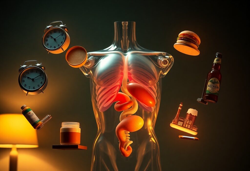
Your everyday behaviors – activity level, food choices, sleep, alcohol and tobacco – accelerate hormonal decline more than chronological age alone. Prolonged sitting, chronic under‑feeding, and disrupted sleep blunt testosterone, GH and IGF‑1 pulses, while excess alcohol and smoking elevate cortisol and impair sex steroid synthesis. Population surveys attribute a large share of between‑person hormone variance to lifestyle, so targeted changes can preserve anabolic tone and metabolic resilience for years.
Sedentary behavior and sarcopenia: loss of anabolic stimulus
When you sit for long stretches your muscles stop sending anabolic signals, driving sarcopenia – muscle mass falls about 3-8% per decade after age 30 and accelerates after 60. Even modest resistance training (2-3 sessions/week, 2-4 sets of 8-12 reps) restores muscle‑derived IGF‑1, increases basal testosterone and improves GH pulsatility within 8-12 weeks, reversing inactivity‑driven hormonal downshifts.
Poor diet and micronutrient deficiencies that impair hormone synthesis
Insufficient protein and micronutrient gaps blunt your hormone factory: intakes below 0.8 g/kg/day favor muscle loss, while older adults typically need 1.2-1.6 g/kg to sustain anabolic signaling. Low 25(OH)D (<20 ng/mL), zinc shortfalls (RDA men 11 mg, women 8 mg) and magnesium deficits impair testosterone, thyroid conversion and steroidogenesis. High‑sugar or chronically restricted diets further raise insulin resistance and suppress sex steroid production.
Practical fixes yield measurable gains: aim for 25-40 g protein per meal and 1.2-1.6 g/kg/day total, test 25(OH)D and target 30-50 ng/mL (often via 1,000-2,000 IU/day individualized by labs), and include zinc‑rich foods (oysters, beef), magnesium (leafy greens, nuts) and occasional Brazil nuts for selenium (~50-90 µg each). Monitor zinc and fat‑soluble vitamin dosing; these adjustments commonly improve testosterone, thyroid markers and energy within weeks to months.
Sleep, circadian and stress-related drivers
Chronic sleep deprivation and reduced anabolic hormone release
If you regularly sleep less than the CDC-recommended 7+ hours, your slow-wave sleep-when most daily growth hormone is released-shrinks and anabolic hormone pulses decline; one study found healthy young men restricted to 5 hours nightly for one week had a 10-15% drop in daytime testosterone. Over months that lowers muscle protein synthesis, impairs recovery, and accelerates sarcopenia and bone loss.
Circadian disruption (shift work, light at night) desynchronizing endocrine rhythms
When your behavioral schedule conflicts with the internal clock-night shifts or bright screens at night-melatonin secretion is suppressed and the phase of cortisol, insulin and reproductive hormone rhythms shifts; research on circadian misalignment shows roughly a 20% reduction in insulin sensitivity and increased nocturnal cortisol, and IARC classifies shift work that disrupts circadian timing as probably carcinogenic (Group 2A).
Mechanistically, light at night-even low intensities around 5-10 lux from streetlight or screens-reduces nocturnal melatonin, which normally interacts with the hypothalamic-pituitary axis and peripheral clocks; this blunting alters LH/FSH timing, impairs glucose homeostasis, and over years is linked to higher rates of metabolic syndrome and some hormone-sensitive cancers in rotating shift cohorts.
Prolonged psychological stress and HPA‑axis overactivity
If you live under chronic stress your HPA axis ramps up baseline cortisol and flattens its diurnal slope, which suppresses GnRH/LH pulsatility and reduces gonadal steroid output; sustained cortisol elevation also promotes visceral fat and insulin resistance, shifting your hormonal milieu toward lower anabolic and higher catabolic signaling that accelerates age-related decline.
At the cellular level, prolonged cortisol exposure downregulates Leydig and granulosa cell steroidogenesis and increases 11β-HSD1 activity in adipose tissue, amplifying local cortisol; longitudinal caregiver and PTSD studies show altered diurnal cortisol profiles with concomitant reductions in DHEA and higher cardiometabolic risk, illustrating how persistent stress accelerates endocrine aging.
Environmental and chemical drivers
Ambient chemicals and pollutants accelerate hormonal decline by accumulating in tissues and disrupting receptor signaling, hormone synthesis, and metabolic clearance; for example, PFAS and PCBs persist for years to decades in your body, altering thyroid and sex-steroid axes and raising metabolic and reproductive risks, while heavy metals like lead and cadmium target gonads and the pituitary, so your lifetime exposure profile matters as much as chronological age.
Endocrine‑disrupting chemicals (BPA, phthalates) interfering with signaling
You encounter BPA from thermal receipts and polycarbonate plastics and phthalates in many personal‑care products and vinyl; both classes can bind estrogen or androgen receptors or block steroidogenic enzymes, and epidemiologic studies link higher urinary BPA or phthalate metabolites to lower testosterone, poorer sperm parameters, and altered thyroid and reproductive hormone patterns in men and women.
Persistent pollutants and heavy metals that impair endocrine organs
PFAS (PFOA/PFOS), PCBs, dioxins and metals like lead, mercury and cadmium bioaccumulate in fat and bone, often with half‑lives measured in years to decades, and they disrupt thyroid transport, steroidogenesis and pancreatic beta‑cell function-consequences that hasten endocrine aging and increase diabetes and infertility risk.
Mechanistically, these mixtures interfere at multiple points: PCBs and dioxins alter hepatic metabolism and deiodinase activity, lowering circulating T4/T3 availability; PFAS compete for binding to transport proteins and can blunt gonadotropin responses; lead and cadmium damage Leydig and ovarian granulosa cells and impair gonadal steroid output. Cohort data-for example, Great Lakes fish‑consuming populations-show PCB exposure correlating with longer time‑to‑pregnancy and reduced sperm quality, and occupational mercury or lead exposure repeatedly links to altered menstrual cycles and reduced testosterone. You can monitor exposure via blood/urine tests (blood lead, serum PFAS, urinary PCB metabolites) when suspecting environmental contributions to hormonal decline.
Metabolic and inflammatory drivers
Visceral adiposity and insulin resistance altering hormone balance
As you accumulate visceral fat, aromatase in VAT converts androgens to estrogens, lowering free testosterone and SHBG; concurrently hyperinsulinemia (HOMA‑IR >2.5) suppresses SHBG and blunts hepatic IGF‑1 production. Visceral adipocytes secrete leptin, resistin and inflammatory cytokines (IL‑6, TNF‑α), promoting central leptin resistance and reducing pulsatile GH release. Clinically this shows as declining libido, muscle loss and impaired glucose control despite stable BMI.
Chronic low‑grade inflammation and immunosenescence impacting hormone action
When you develop inflammaging, circulating IL‑6 and CRP (CRP >3 mg/L) rise and senescent immune cells release SASP factors that desensitize insulin and GH receptors, lower IGF‑1 and blunt gonadotropin signaling. Thymic involution and reduced T‑cell diversity shift cytokine profiles toward TNF‑α and IL‑1β, reinforcing HPA axis activation and higher cortisol rhythms that antagonize sex steroids and anabolic hormones, accelerating sarcopenia and metabolic decline.
Mechanistically, the SASP from p16+/p21+ senescent cells-rich in IL‑6, IL‑8 and MMPs-alters hypothalamic signaling and impairs pituitary responsiveness, so you see blunted nocturnal GH peaks and lower DHEA. Interventions that reduce inflammation-regular resistance training, modest weight loss (5-10%), Mediterranean‑style diets or targeted senolytic approaches in preclinical studies-restore hormonal responsiveness and lower IL‑6/CRP, improving muscle mass and metabolic markers.
Mechanisms, clinical consequences and intervention priorities
Your hormonal decline reflects interacting processes: progressive hypothalamic-pituitary attenuation, increased adipose aromatase activity, chronic inflammation and altered hepatic clearance. Testosterone in men falls roughly 1% per year after age 30, while DHEA and growth hormone drop substantially, driving sarcopenia, visceral fat gain, osteoporosis and mood/cognitive changes. Prioritize screening high-risk patients (obesity, diabetes, sleep apnea, chemo exposure), target reversible drivers first, and reserve hormone replacement for symptomatic individuals with confirmed biochemical deficiency.
How these drivers converge on hormone production, receptor sensitivity and clearance
Inflammation (IL-6, TNF-α) and insulin resistance suppress hypothalamic GnRH and pituitary output, while adiposity increases aromatase, converting androgens to estrogens and raising SHBG, which lowers free hormone fractions. Environmental endocrine disruptors impair receptor signaling and hepatic enzyme induction speeds steroid clearance. As a result, you may have normal total hormone levels but reduced tissue exposure from receptor downregulation and increased binding protein activity, amplifying functional deficiency.
Screening, lifestyle and medical strategies to slow hormonal decline
Start with targeted screening: morning total and free testosterone, SHBG, fasting glucose, lipids and sleep assessment in men; FSH/LH and symptom inventory in women. You should pursue weight loss (5-10% bodyweight), resistance training 2-3×/week, 150 min/wk aerobic exercise, 7-9 hours sleep and treat sleep apnea-each improves insulin sensitivity and hormone profiles. Consider metformin/GLP-1 for metabolic drivers and hormone therapy only after confirming deficiency and weighing risks.
For implementation, screen symptomatic high-risk adults annually and confirm low testosterone with two morning measurements (total <300 ng/dL). If you start testosterone, check hematocrit at 3 and 6 months, then yearly; stop or adjust if hematocrit >54% or PSA rises substantially. Use DEXA for fracture risk (women ≥65, men ≥70 or earlier with risk factors). Document symptom response and biochemical targets to guide dose titration and reassessment every 6-12 months.
Summing up
Hence you should understand that lifestyle factors, sleep, stress, environmental toxins, inflammation, medications, weight, exercise habits and nutrient deficiencies each speed hormonal decline; by addressing these nine hidden drivers through targeted changes and medical guidance you can slow hormonal aging, preserve function, and improve your energy and long-term health.