Healing begins when you understand how brain waves shape hormonal balance, sleep quality, and tissue repair; this post outlines five evidence-based mechanisms by which specific brain states-delta, theta, alpha, beta, and gamma-modulate cortisol, melatonin, growth hormone, autonomic tone, and inflammatory responses. You will learn practical implications for sleep hygiene, stress management, therapeutic interventions, and recovery strategies to harness your brain states for better health and healing.
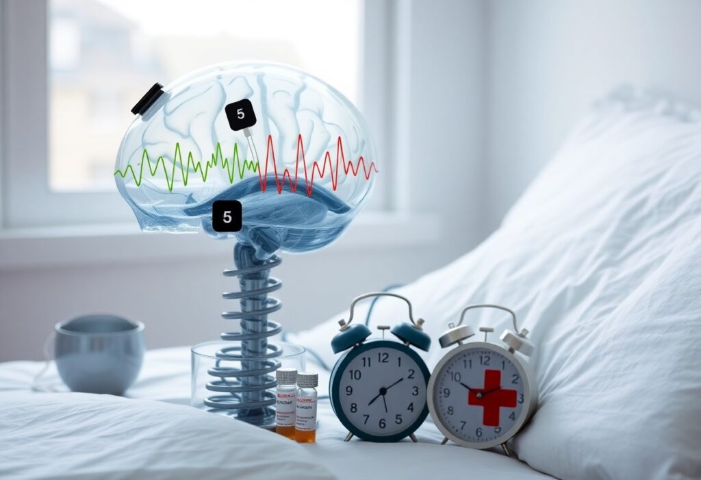
Brain Waves 101
You can think of brain waves as rhythmic electrical patterns produced by synchronized neuronal populations; their frequency and amplitude shape your arousal, memory consolidation, and hormonal signaling via thalamocortical and hypothalamic pathways. Measured in hertz, these oscillations gate neurotransmitter release, influence melatonin and cortisol timing, and set the neural context for healing and sleep architecture.
Frequency bands and functional roles (delta, theta, alpha, beta, gamma)
Delta (0.5-4 Hz) dominates deep N3 sleep and restorative growth processes, theta (4-8 Hz) links to hippocampal memory replay and creativity, alpha (8-12 Hz) marks relaxed wakefulness and inhibition of irrelevant input, beta (12-30 Hz) supports focused attention and motor control, and gamma (≈30-100 Hz) coordinates binding and high-level cognition across networks.
Measurement and state transitions (EEG, wearables, biomarkers)
Scalp EEG using the 10-20 montage remains the clinical standard for spectral and event-related analyses; consumer wearables (EEG headbands, HRV rings, EDA patches) provide continuous proxies. State transitions occur over milliseconds for microstates, minutes for sleep-stage shifts, and ~90-minute REM cycles. You should expect sampling ≥250 Hz to capture gamma activity and anticipate motion and electrode impedance as common confounds.
Power spectral density, coherence, and event-related potentials (like P300 latency ~300 ms) are the analytic tools you use to quantify state. Peripheral biomarkers-cortisol’s awakening peak ~30-45 minutes post-wake, melatonin onset, HRV (RMSSD)-correlate with EEG states. In validation studies consumer headbands and sleep trackers often reach ~80-90% agreement with PSG for broad sleep stages, but you must account for lower spatial resolution, motion artifacts, and dry-electrode impedance trade-offs when interpreting results.
Five Proven Ways Brain States Influence Physiology
You feel the effects across five pathways: endocrine modulation, sleep-driven cellular repair, autonomic balance, pain and wound-healing trajectories, and neuroimmune signaling. Delta-dominant slow-wave sleep drives growth hormone release and glymphatic clearance; theta/alpha states enhance parasympathetic recovery and tissue perfusion; beta/gamma arousal spikes HPA output and sympathetic tone. Experimental entrainment of slow oscillations increases restorative markers, while chronic cortical hyperarousal raises evening cortisol and blunts vaccine and wound-healing responses in clinical studies.
Endocrine modulation – cortisol, melatonin, and sex hormones
Your brain-state shifts directly alter hormone timing and amplitude: arousal-related beta/gamma bursts trigger rapid HPA activation and cortisol pulses, while sustained cortical quiet promotes melatonin secretion and a low-cortisol trough. Sleep loss and fragmented slow-wave sleep lower nocturnal testosterone-controlled trials report ~10-15% drops after successive five-hour nights-and chronic arousal flattens cortisol rhythm, worsening metabolic and reproductive outcomes.
Sleep architecture and cellular repair – growth hormone, memory consolidation, immune signaling
Your slow-wave sleep (SWS) is where most growth hormone is released and glymphatic clearance accelerates: roughly 70% of daily GH secretion occurs during early-night SWS, and rodent work shows interstitial space expands ~60% with roughly twofold CSF-mediated clearance of metabolites. Concurrently, slow oscillations and 12-15 Hz spindles coordinate hippocampo-cortical transfer for declarative memory and modulate immune signaling that affects vaccine responses and inflammatory markers.
SWS predominates in the first third of the night and typically comprises ~20-25% of total sleep in young adults, declining markedly with age; that timing concentrates GH pulses and synaptic downscaling. Techniques that boost slow-wave power-auditory closed-loop stimulation or transcranial stimulation-have increased slow oscillation amplitude by ~30-40% and improved word-pair retention by ~15-25% in lab trials. At the same time, impaired SWS correlates with higher amyloid burden and elevated IL-6/CRP, linking reduced overnight clearance and heightened inflammation to long-term neurodegenerative and immune risk.
Brain States and Healing
Neuroimmune interactions and inflammation control
When you shift into calm alpha or theta states your vagal tone increases and engages the cholinergic anti-inflammatory pathway: acetylcholine acting on α7 nicotinic receptors reduces TNF‑α and IL‑6 release from macrophages. Vagus nerve stimulation and HRV biofeedback have been shown to lower systemic inflammation in clinical and preclinical studies, and simple practices like 5-20 minutes of paced breathing done daily can produce measurable HRV improvements and downstream reductions in inflammatory markers over weeks.
Neuroplasticity, pain modulation, and recovery
Theta/alpha entrainment and targeted stimulation enhance synaptic plasticity and pain gating by elevating BDNF and engaging descending inhibitory circuits; you can use tDCS (1-2 mA) or rTMS (commonly 10 Hz) to reduce neuropathic pain in trials and to prime motor cortex for rehab. Applying these methods alongside behavioral therapy produces larger functional gains than either alone, accelerating recovery after injury.
Timing and dose matter: applying stimulation immediately before or during task‑specific practice yields LTP‑like changes and cortical map reorganization, especially within the first 3 months post‑stroke when plasticity windows peak. Aim for repeated courses-typically 5-20 sessions over 1-4 weeks-paired with intensive, goal‑directed training to maximize strength, gait, and pain threshold improvements.
Sleep, Disorders, and Clinical Implications
You encounter distinct EEG fingerprints across sleep disorders: chronic insomnia (affecting ~10% of adults) shows elevated beta/gamma and reduced delta during NREM, narcolepsy features REM intrusion tied to low orexin, and PTSD presents fragmented REM with heightened high-frequency power and nightmare density. Clinically, these signatures predict response to interventions-CBT-I, targeted pharmacotherapy, neurofeedback, or slow-wave enhancement-and guide monitoring of recovery and relapse risk using ambulatory EEG or polysomnography.
How altered brain waves produce insomnia, hypersomnia, and PTSD-related patterns
You develop insomnia when persistent high-frequency beta/gamma activity prevents sleep-onset and suppresses slow-wave generation; studies report 10-30% higher high-frequency power in insomniacs during NREM. Hypersomnia and narcolepsy arise from abnormal slow-wave/REM regulation-narcolepsy shows REM intrusions due to orexin loss. In PTSD, limbic hyperactivation increases phasic high-frequency bursts and REM fragmentation, producing nightmare consolidation and impaired restorative slow-wave sleep.
Downstream hormonal and metabolic consequences
You experience hormonal shifts when slow-wave loss alters endocrine timing: growth hormone release (concentrated in the first 90 minutes of SWS) drops, nocturnal cortisol becomes elevated or flattened, and one night of sleep restriction can reduce insulin sensitivity by roughly 20-30%. Appetite-regulating hormones flip-leptin falls while ghrelin rises-raising caloric intake and metabolic risk, and proinflammatory cytokines like IL-6 and TNF-α increase with chronic disruption.
You can see these changes translate into clinical endpoints: chronic sleep-wave disruption associates with higher fasting glucose, increased waist circumference, and greater odds of metabolic syndrome in cohort studies. Interventions that boost slow-wave activity-CBT-I, timed exercise, or phase-locked auditory stimulation-have improved GH secretion, reduced nocturnal cortisol excursions, and partially restored insulin sensitivity in pilot trials, offering measurable metabolic benefit when EEG patterns normalize.

Modulating Brain States – Evidence-Based Interventions
Behavioral and environmental strategies (sleep hygiene, light exposure, exercise)
You can reshape sleep and hormones with targeted behavioral changes: fix your wake time, avoid screens 60 minutes before bed, and cut caffeine about 6 hours before sleep. Morning bright light (~10,000 lux for 20-30 minutes within 30 minutes of waking) shifts circadian phase and suppresses melatonin. Aim for 150 minutes/week moderate or 75 minutes vigorous exercise; earlier aerobic or resistance training boosts growth-hormone pulses and slow-wave sleep. Keep your bedroom around 16-19°C for better sleep consolidation.
Direct modulation (meditation, neurofeedback, binaural beats, noninvasive stimulation)
You can directly alter rhythms with practices and devices that target specific frequencies: eight-week MBSR or focused-attention training increases frontal alpha/theta and reduces stress, while neurofeedback (theta/beta, SMR) yields medium effects for attention and insomnia after 20-40 sessions. Binaural beats in delta/theta ranges show small reductions in anxiety and improved sleep in short trials. Noninvasive stimulation-tDCS (1-2 mA, ~20 minutes) and rTMS (10 Hz or iTBS to left DLPFC)-modulates cortical excitability and mood.
Practical dosing matters: meditate 10-30 minutes daily or enroll in an 8-week course for measurable structural and EEG changes; expect neurofeedback to require 20-40 sessions (2-3×/week) for lasting gains. For binaural beats use headphones, delta 0.5-4 Hz for sleep or theta 4-8 Hz for relaxation, 15-45 minutes at low volume. If you consider tDCS or rTMS, work with a clinician-typical tDCS montages use anodal left DLPFC at 1-2 mA for 20 minutes, while rTMS often runs 5 days/week for 4-6 weeks for antidepressant effects.
Monitoring and Practical Guidance
You should pair objective tracking, targeted testing, and staged interventions to turn brain-state insights into measurable outcomes. Use wearables and home EEG to quantify sleep architecture and HRV, add diurnal hormone panels (4‑point salivary cortisol, DLMO melatonin, timed sex‑hormone draws), and adopt single‑variable changes with weekly monitoring so you can attribute effects and adjust within a 4-8 week window.
Tracking brain states and sleep – wearables, home EEG, hormone testing
You can use consumer wearables (Oura, Apple Watch, Whoop) for sleep staging, HRV and respiratory rate; home EEG devices (Muse S, Dreem 2, OpenBCI) capture alpha/theta transitions and nightly sleep scoring. Order a 4‑point salivary cortisol (wake, +30 min, midday, bedtime) and a DLMO salivary melatonin test to map circadian phase, and schedule sex‑hormone blood draws to specific cycle days for accurate comparison.
When to seek professional assessment and how to integrate interventions safely
You should seek specialist input if insomnia or hypersomnia persists beyond 3 months with daytime impairment, if home EEG suggests epileptiform activity, or if hormone panels show values outside reference ranges (e.g., blunted cortisol awakening response). Coordinate with a sleep physician, neurologist, or endocrinologist; introduce one intervention at a time, document baseline metrics, check for drug interactions, and monitor objective responses weekly.
You begin with a structured assessment: keep a two‑week sleep diary plus actigraphy, obtain baseline labs (TSH, fasting AM cortisol, 4‑point salivary cortisol, salivary DLMO, estradiol/testosterone timed to cycle), and pursue diagnostic testing when indicated (overnight polysomnography for suspected OSA, EEG for seizures). When you integrate treatments, phase them-start nonpharmacologic options first (bright‑light therapy 10,000 lux, 20-30 minutes each morning; CBT‑I for 6-8 weekly sessions), then add low‑dose melatonin (0.5-1 mg, 1-2 hours before target sleep) if needed; reserve sedatives or hormone replacement for specialist oversight. You should recheck objective metrics at 6-12 weeks and labs at 8-12 weeks, and escalate care immediately if you develop suicidal thoughts, syncope, chest pain, or new focal neurological signs.
Final Words
Following this, you can apply insights about alpha, beta, theta, delta and gamma activity to influence your hormones, improve sleep quality, speed healing, reduce inflammation, and strengthen recovery, guiding practical strategies that support your long-term brain-body health.