Repair systems that preserve cellular integrity are among the first targets of oxidative stress; in this post you’ll see how oxidative damage impairs DNA repair, protein quality control (proteostasis), mitochondrial maintenance, lipid membrane repair, redox signaling and autophagy, and what practical strategies you can use to support your cells’ repair capacity. By understanding these mechanisms you’ll be better equipped to evaluate interventions that restore cellular resilience and reduce long-term dysfunction.
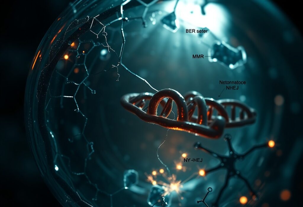
Base‑excision DNA repair (BER)
BER removes small, non‑helix‑distorting lesions-most commonly oxidized bases like 8‑oxoguanine (8‑oxoG), deaminated bases, and abasic (AP) sites-by converting each lesion into a single‑strand break that is then processed and sealed. When oxidative load rises to 10^4-10^5 lesions per cell per day, BER throughput determines whether your genome accumulates mutations or maintains fidelity during replication.
Core functions and key enzymes (OGG1, APE1, Pol β, XRCC1)
OGG1 recognizes and excises 8‑oxoG via glycosylase/AP‑lyase activity; APE1 incises AP sites and also provides redox signaling; Pol β fills single‑nucleotide gaps and removes the 5’‑dRP moiety; XRCC1 scaffolds Pol β, LigIII and end‑processing factors to coordinate handoffs and increase repair efficiency. You depend on precise timing and protein-protein contacts to avoid persistent SSBs that become toxic.
How oxidative lesions and enzyme oxidation disable BER
Excess 8‑oxoG, clustered oxidations and AP site accumulation overload BER, producing repair intermediates that stall replication and elevate double‑strand break risk. At the same time, ROS/RNS modify repair enzymes-sulfenylation, S‑glutathionylation or nitrosylation of active cysteines-reducing OGG1 excision, APE1 incision, and Pol β polymerase/lyase activities, so your repair capacity collapses under sustained oxidative pressure.
Mechanistically, oxidation can trap OGG1 on DNA or change its substrate specificity, while APE1 redox changes decouple its endonuclease and transcriptional roles. You also face NAD+ depletion from PARP1 hyperactivation at clustered SSBs, which impairs XRCC1 recruitment and LigIII sealing; chronic oxidation promotes ubiquitin‑mediated degradation of modified Pol β, further lowering single‑nucleotide BER output and increasing mutagenesis in inflamed or aged tissues.
Mitochondrial quality control and mitophagy
You rely on mitochondrial quality control to remove ROS-damaged organelles before they poison your cell; about 80-90% of intracellular ROS originates in mitochondria, so fission/fusion, mitophagy and biogenesis are constantly balancing turnover. In tissues with high energy demand like heart and brain, impaired clearance accelerates dysfunction-PINK1/Parkin pathway defects in familial Parkinson’s provide a concrete example of how failed mitophagy leads to accumulation of depolarized, ROS-producing mitochondria.
Mitochondrial dynamics, mitophagy and mtDNA maintenance
You modulate mitochondrial health via DRP1-driven fission and MFN1/2- and OPA1-mediated fusion, which segregate and dilute damage. Human mtDNA is ~16,569 bases and copy number ranges from ~100 in leukocytes to thousands in muscle; fusion helps maintain functional mtDNA pools while selective mitophagy clears heavily mutated genomes, limiting deletions such as the 4,977‑bp “common deletion” seen in aging tissues.
ROS‑driven mitochondrial dysfunction and impaired clearance
You face a feed‑forward loop when ROS oxidize lipids, proteins and mtDNA: oxidized cardiolipin can signal for mitophagy, yet excessive ROS also inactivate Parkin/PINK1 function and proteostasis components, reducing clearance. In neurodegenerative and metabolic disease models, this imbalance produces sustained mitochondrial depolarization, higher superoxide/H2O2 emission and progressive loss of respiratory capacity.
Mechanistically, ROS modify Parkin cysteines and impair its E3 ligase activity, while mtDNA damage-point mutations and deletions-lowers complex I/III efficiency and increases ROS output. You can detect these changes clinically via decreased citrate synthase activity and altered mtDNA copy number; interventions like exercise, NAD+ precursors and mitophagy inducers (e.g., urolithin A in preclinical and early human studies) restore turnover and break the ROS-amplifying cycle.
Proteostasis: chaperones, ubiquitin-proteasome and autophagy
When oxidative damage accumulates, your proteostasis network shifts between refolding, tagging, and disposal to limit toxicity: Hsp70/Hsp40 and Hsp90 cycles attempt refolding using ATP-dependent clamps, E1-E2-E3 ubiquitination cascades (including CHIP and Parkin) mark persistent misfolded proteins with K48 chains for the 26S proteasome (~2.5 MDa), and macroautophagy receptors like p62/SQSTM1 and NBR1 sequester larger oligomers for lysosomal clearance.
Protein refolding, ubiquitination and degradation pathways
Chaperone systems (Hsp70/Hsp40/Hsp90) initially bind exposed hydrophobic patches to promote ATP-driven refolding, while folding failures are routed by cochaperones (BAGs, CHIP) that recruit E3 ligases to build polyubiquitin chains; thereafter the 26S proteasome recognizes K48-linked chains and unfolds substrates for the 20S core, and when soluble clearance is overwhelmed your cells switch to selective autophagy mediated by p62 and LC3.
Oxidation‑induced misfolding, proteasome inhibition and aggregation
Oxidative modifications – protein carbonylation, cysteine sulfenylation, methionine oxidation and crosslinks – destabilize native folds so proteins expose sticky regions that seed oligomers; concurrently, the ATP‑dependent 26S proteasome is functionally impaired under ROS and ATP depletion, shifting reliance to ubiquitin‑independent 20S degradation and autophagy, which explains why you see ubiquitin‑positive inclusions in conditions like Alzheimer’s and Parkinson’s.
Mechanistically, oxidized polypeptides resist chaperone‑mediated unfolding and often form β‑rich oligomers that sterically block proteasomal entry or bind and sequester 19S subunits; oxidative stress also promotes 26S disassembly and lowers chymotrypsin‑like activity, forcing a proteolytic triage-20S proteasome handles soluble oxidized proteins while p62‑dependent macroautophagy clears large, crosslinked aggregates. For example, dopamine oxidation in nigral neurons generates quinones that modify α‑synuclein and accelerate Lewy body formation, and mutations that impair Parkin or autophagy genes (e.g., PINK1, ATG5) heighten aggregation and neurodegeneration in model systems, so your cell’s outcome depends on the balance between these competing repair and disposal routes.
Lipid repair and membrane integrity
Your membranes, rich in polyunsaturated phospholipids, require constant repair: phospholipase A2 removes oxidized acyl chains, lysophospholipid acyltransferases (LPCAT3) and ACSL4 re-esterify repaired lipids, while GPX4 detoxifies hydroperoxides. When repair is overwhelmed, leakage, ion dysregulation and signaling collapse follow within hours; in ischemia-reperfusion and cancer models, modulating these enzymes shifts cell survival and membrane stability.
Phospholipid repair, GPX4 and prevention of lipid peroxidation
GPX4, a selenoenzyme, reduces phospholipid hydroperoxides to alcohols using two glutathione (GSH) molecules, preventing chain reactions; genetic GPX4 ablation is embryonically lethal in mice and neuron-specific loss causes rapid degeneration. When you deplete GSH or inhibit GPX4, lipid peroxides accumulate and ferroptosis ensues, yet lipophilic antioxidants like ferrostatin-1 or liproxstatin-1 can restore membrane function in cell assays.
Membrane disruption, lipid peroxidation cascades and ferroptosis
If iron-catalyzed reactions initiate peroxidation of arachidonoyl/adrenoyl chains, propagation through bis-allylic hydrogens creates lipid radicals and breakdown products (4-HNE, MDA) that crosslink proteins and increase permeability. You’ll see condensed mitochondria and loss of plasma membrane integrity in ferroptosis; preventing it with iron chelators (deferoxamine) or lipophilic radical scavengers underscores the membrane-centered mechanism of this death pathway.
Mechanistically, ACSL4 and LPCAT3 channel PUFAs into phosphatidylethanolamines (PE-AA/PE-AdA), which lipoxygenases (ALOX5/15) preferentially oxidize to PE-OOH species; accumulation of low-micromolar oxidized PEs suffices to trigger ferroptosis in cultured cells. Therapies that lower labile iron, inhibit ACSL4, or restore GPX4 activity shift thresholds for membrane collapse, so you can exploit ferroptosis against therapy-resistant tumors but must avoid collateral damage in stroke models.
Antioxidant enzyme systems and redox signaling
You rely on coordinated antioxidant enzymes to keep signaling thiols responsive while clearing reactive species; SOD isoforms convert O2•− to H2O2, catalase and peroxisomal enzymes decompose H2O2, and the GSH/Trx systems buffer redox couples so that your GSH:GSSG ratio (normally >100:1) and NADPH supply sustain reversible thiol-based signaling rather than protein destruction.
SOD, catalase, glutathione and thioredoxin systems in redox balance
You depend on SOD1 (Cu/Zn, cytosol), SOD2 (Mn, mitochondria) and SOD3 (extracellular) for rapid O2•− dismutation (~10^9 M^-1s^-1). Catalase in peroxisomes and peroxiredoxins remove H2O2; glutathione (1-10 mM intracellular) plus glutaredoxin manage S‑glutathionylation, while thioredoxin and thioredoxin reductase (NADPH-dependent) restore protein disulfides, supporting processes like ribonucleotide reductase and redox signaling fidelity.
Enzyme inactivation, redox signaling breakdown and feedback failure
You see failure when high oxidant flux irreversibly modifies active-site cysteines (sulfonic acid), carbonylates enzymes, or hyperoxidizes peroxiredoxin; clinically this appears as GSH depletion in acetaminophen liver injury and mutant SOD1 aggregation in ~10-20% of familial ALS cases, both examples where antioxidant systems collapse and signaling feedback loops stop functioning.
Delving deeper, reversible cysteine sulfenylation (SOH) acts as a signaling switch but progresses to sulfinic (SO2H) or sulfonic (SO3H) states under sustained H2O2; sulfinic peroxiredoxin can be recycled by ATP-dependent sulfiredoxin, whereas sulfonylation and carbonylation are largely irreversible. You need NADPH (from G6PD and the pentose phosphate pathway) to drive thioredoxin and glutathione recycling; G6PD deficiency, for example, lowers NADPH and predisposes red cells to hemolysis under oxidant stress. Proteasome overload by oxidized proteins further impairs clearance, promoting aggregates seen in neurodegeneration and breaking the feedback that normally restores redox homeostasis.
Double‑strand break repair and DNA damage signaling
You depend on tightly coordinated DSB repair and signaling: sensors (MRN, RPA) recruit ATM/ATR, which phosphorylate H2AX and checkpoint kinases to remodel chromatin and pause the cell cycle, while NHEJ and HR execute repair; oxidative stress perturbs sensors, signaling amplitude and repair choice, shifting outcomes toward mutagenesis, chromosome rearrangements and persistent DDR foci.
NHEJ/HR pathways and ATM/ATR checkpoint activation
You use NHEJ (Ku70/Ku80, DNA‑PKcs, XRCC4-Ligase IV) for rapid end‑joining in G0/G1 and HR (resection by MRE11/CtIP, RAD51 filament formation, BRCA1/2, PALB2) in S/G2 for high‑fidelity repair. ATM is activated at DSBs via MRN to generate γ‑H2AX over megabase domains and activate CHK2; ATR senses RPA‑coated ssDNA at stalled forks and phosphorylates CHK1 to coordinate replication restart and fork protection.
Oxidative impairment of repair proteins, genomic instability and senescence
You see oxidative modifications-cysteine oxidation, carbonylation and 4‑HNE adducts-directly impair Ku, DNA‑PKcs, BRCA1/2 and RAD51, blunting end‑joining fidelity and strand invasion. As signaling falters, you accumulate unrepaired DSBs, micronuclei and translocations, and cells shift into p21/p16‑mediated senescence with a pro‑inflammatory SASP that amplifies tissue dysfunction.
Further, oxidative hits trigger proteasomal clearance and mislocalization of repair factors, and PARP1 hyperactivation can deplete NAD+ to impede ATP‑dependent chromatin remodelers and sirtuin‑mediated repair regulation. Telomeres-rich in guanines-preferentially form 8‑oxoG, accelerating shortening and end‑to‑end fusions; in culture, these combined effects measurably raise mutation frequency and accelerate senescence markers within a few population doublings.
To wrap up
Summing up, oxidative stress attacks your cellular resilience by disrupting six repair pathways first: DNA repair, proteostasis, mitochondrial quality control, lipid repair, antioxidant recycling, and inflammation resolution; preserving and restoring these systems through diet, exercise, targeted supplementation, and medical intervention helps you directly protect genome integrity, protein function, energy production, membrane stability, redox balance, and immune homeostasis.