With common blood tests often missing low-grade inflammation, you need to watch for signs at the cellular level that signal trouble: persistent fatigue, brain fog, unexplained weight changes, chronic muscle or joint pain, frequent infections, poor wound healing, digestive dysfunction, skin rashes, and mood disturbances. Understanding these nine warning signs helps you advocate for targeted testing and early interventions to protect long-term health.
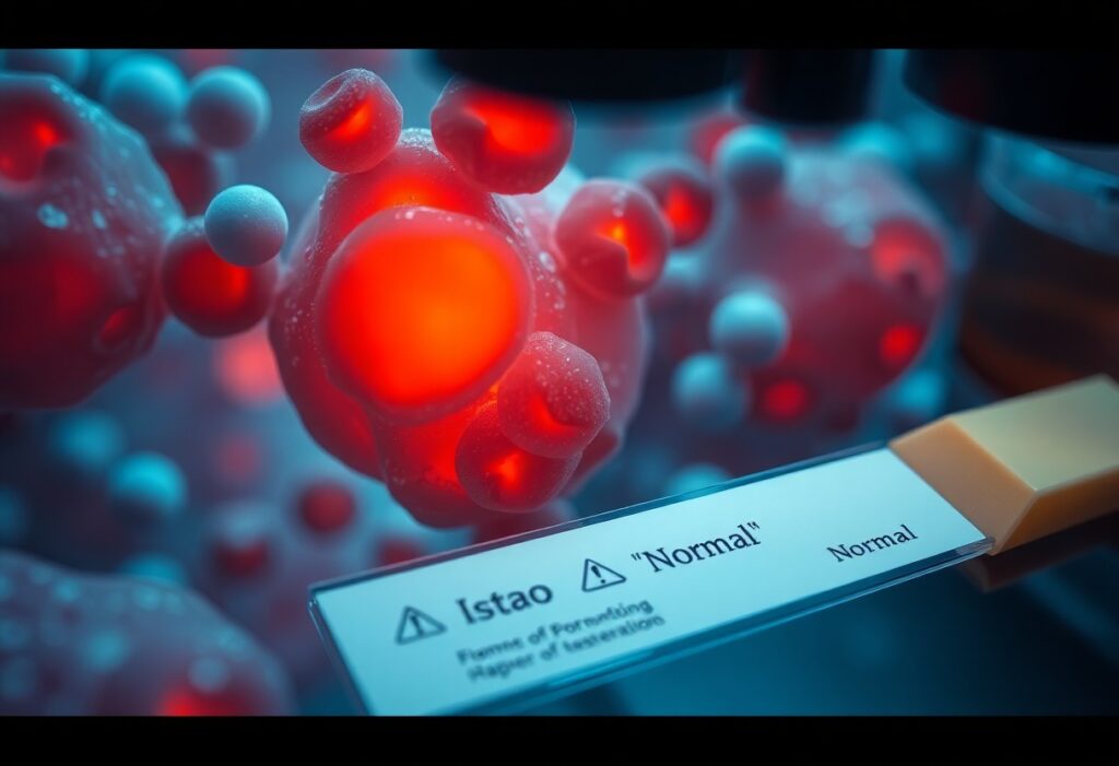
Cellular inflammation explained
At the cellular level, inflammation is a coordinated response: sensing by pattern-recognition receptors, activation of signaling hubs like NF-κB and NLRP3, release of cytokines (IL-1β, IL-6, TNF-α), mitochondrial ROS production, and shifts in metabolic programs that rewire gene expression and proteostasis; you can have this activated locally in tissue or persist subtly for years, driving dysfunction long before systemic tests flag a problem.
Definition and core cellular mechanisms
Innate sensors (TLRs, NLRs, cGAS-STING) detect PAMPs/DAMPs and trigger kinase cascades (MAPK, IKK) that activate transcription factors such as NF-κB and IRFs; you should note that inflammasome-driven caspase-1 cleavage yields IL-1β/IL-18, while mitochondrial dysfunction, impaired autophagy, and altered NAD+/sirtuin signaling amplify a feed-forward inflammatory state at the single-cell level.
Acute vs. chronic: localized, low‑grade and systemic effects
Acute inflammation lasts hours-days, recruits neutrophils, and promotes repair, whereas chronic low-grade inflammation can persist for months-years with macrophage predominance, fibroblast activation, and subtle systemic signals-high-sensitivity CRP often sits in the 1-3 mg/L range for low-grade inflammation while severe systemic inflammation pushes CRP >10 mg/L; you may feel fatigue or metabolic shifts despite normal routine panels.
For example, adipose tissue in obesity shows expanded M1-like macrophages that release IL-6 and TNF-α, promoting hepatic insulin resistance and modestly elevating hs-CRP; similarly, senescent cells secrete a SASP rich in IL-6/IL-8 that sustains local inflammation, and NLRP3 activation by cholesterol crystals or urate can link local deposits to systemic vascular inflammation-these mechanisms explain why tissue-level inflammation often precedes clear blood-test abnormalities.
Why routine tests can appear “normal”
Your blood work can look unremarkable despite active cellular inflammation because most routine assays sample systemic spillover, not tissue microenvironments. Standard CRP and ESR detect broad, high-amplitude acute responses-values under common cutoffs (CRP <10 mg/L) will be reported as normal even when low‑grade or localized cytokine activity is present. Timing matters: CRP has a ~19‑hour half‑life, so short spikes can be missed, and up to half of patients with certain inflammatory disorders show normal CRP despite symptoms.
Limitations of common markers (CRP, ESR, standard panels)
You rely on CRP and ESR because labs report them routinely, but both have blind spots: CRP often needs >10 mg/L to flag, while high‑sensitivity CRP (hs‑CRP) reveals 0.3-10 mg/L ranges used for cardiovascular risk rather than tissue inflammation. ESR is affected by age, sex, anemia and rises slowly. Consequently you can have active autoimmune flares, localized infections or early disease with normal standard panels; clinicians miss inflammatory signals in roughly 30-50% of some conditions.
Compartmentalized, transient and low‑grade inflammation patterns
You should expect three patterns that evade routine tests: compartmentalized inflammation confined to synovium, gut or adipose tissue that doesn’t spill into blood; transient spikes tied to infection, exercise or flare that resolve within 24-72 hours and are missed by timing; and low‑grade chronic inflammation with CRP in the 0.5-3 mg/L range that blends into “normal” lab noise despite driving symptoms and long‑term risk.
You can detect compartmentalized inflammation by targeting the affected compartment: fecal calprotectin for gut, synovial fluid cytokines or ultrasound for joints, and epicardial/adipose imaging or adipokine panels for metabolic inflammation. When symptoms are intermittent, ask for timed sampling or repeat tests 24-72 hours after a flare. Use hs‑CRP, IL‑6 or targeted biomarkers to unmask low‑grade signals that standard panels miss.
Compartmentalized, transient & low‑grade inflammation – quick reference
| Pattern | How it evades routine tests and practical alternatives |
| Compartmentalized (gut, joint, brain) | Local cytokines stay in tissue; blood CRP/ESR often normal. Use fecal calprotectin, synovial fluid analysis, MRI/ultrasound or tissue‑specific biomarkers. |
| Transient (post‑viral, exertional, flare) | Short CRP spikes clear in ~24-48 hours. Time sampling to symptom onset, repeat tests, or use serial hs‑CRP measurements. |
| Low‑grade chronic (metabolic, adipose) | CRP often 0.5-3 mg/L and labeled normal; consider hs‑CRP, IL‑6, ferritin patterns, insulin resistance markers, and targeted imaging. |
Nine warning signs your cells are inflamed
Signs 1-5: fatigue, unexplained pain, digestive issues, sleep disruption, skin changes
You may feel persistent fatigue that doesn’t lift after 7-8 hours of sleep, or diffuse aches and joint stiffness that resist NSAIDs; about 30-50% of people with low-grade inflammation report musculoskeletal pain. Your digestion can show bloating, IBS-like changes, or food sensitivities, while sleep becomes fragmented with frequent awakenings. Skin often signals inflammation too-eczema flares, new acne, or psoriasis worsening can be early clues that your cells are inflamed.
Signs 6-9: brain fog/mood changes, recurrent infections, poor wound healing, metabolic shifts
You might notice brain fog-slower thinking, poor concentration-or mood changes like increased anxiety or low mood linked to inflammatory cytokines. Recurrent infections that outpace peers, wounds that take weeks instead of days to close, and metabolic shifts such as unexplained abdominal weight gain or rising fasting glucose (e.g., from 90 toward 100-110 mg/dL) round out the later signs.
Inflammatory mediators like IL-6 and TNF-α can cross the blood-brain barrier and disrupt neurotransmitter balance, explaining cognitive and mood symptoms; similarly, chronic cytokine exposure impairs neutrophil and macrophage function, increasing infection risk. Collagen synthesis slows under sustained inflammation, often doubling healing time in clinical observations. Metabolically, inflammation promotes serine phosphorylation of insulin receptor substrates, driving insulin resistance that shows up as rising fasting glucose, higher triglycerides, and stubborn visceral fat in case reports.
Clinical clues, patterns and red flags
Symptom clusters and onset patterns that suggest cellular inflammation
You should pay attention when symptoms span 2+ organ systems – for example persistent fatigue, brain fog, sleep disturbance plus gut pain or headaches – especially if onset followed an infection or toxin exposure within 2-6 weeks. Day-to-day variability with post-exertional worsening, symptoms lasting >3 months, and disproportionate functional decline (work or exercise capacity down by 30-50%) are classic patterns that point toward low-grade cellular inflammation despite normal routine labs.
When history and exam outweigh normal labs
You must act on a consistent history or objective exam findings even when CRP, ESR and routine panels are normal. Examples include progressive morning stiffness >30 minutes, focal neurologic deficits, joint swelling, orthostatic drop ≥20 mmHg systolic or heart-rate rise ≥30 bpm, or unexplained weight loss >5% in 6 months. Those signs increase pre-test probability enough to pursue advanced testing or specialist referral rather than dismissing symptoms as “normal labs.”
In practice this means ordering targeted tests (hs-CRP, ferritin, ANA, cytokine panels, autonomic testing, skin biopsy for small-fibre neuropathy) or a timed therapeutic trial when exam and timeline are convincing. Case example: a 38-year-old with post-infectious brain fog and orthostatic intolerance had normal ESR/CRP but tilt-table confirmed POTS and small-fibre neuropathy was later shown on biopsy; early referral changed management and improved function within months.
Tests and advanced biomarkers to consider
-
hs‑CRP (high‑sensitivity C‑reactive protein)
What it measures Clinical insight / when to use Low‑grade systemic inflammation via liver acute‑phase response Use to stratify CV risk: <1 mg/L low, 1-3 mg/L moderate, >3 mg/L higher risk; serial rises over months suggest ongoing inflammation despite normal routine labs. -
Cytokine panels (IL‑6, TNF‑α, IL‑1β)
What it measures Clinical insight / when to use Circulating pro‑inflammatory signaling molecules Order when unexplained symptoms persist; IL‑6 elevations (eg, 5-10 pg/mL) can appear when CRP is borderline; marked values (>10-20 pg/mL) warrant specialty referral. -
Oxidative stress markers (urinary 8‑OHdG, F2‑isoprostanes)
What it measures Clinical insight / when to use Cellular DNA/RNA and lipid oxidation products Helpful in patients with fatigue, neuro symptoms, or environmental exposures; elevated 8‑OHdG or F2‑isoprostanes point to mitochondrial/oxidative injury driving microinflammation. -
Microinflammation panels (GlycA, fibrinogen, complement C3)
What it measures Clinical insight / when to use Integrated glycoprotein acetylation and acute‑phase proteins GlycA from NMR correlates with long‑term inflammatory burden and CV events; use when standard markers are normal but clinical suspicion remains high. -
Cellular activation markers (sCD14, sCD163, monocyte subsets)
What it measures Clinical insight / when to use Monocyte/macrophage activation and innate immune status Elevations suggest chronic innate immune activation (eg, persistent infection, metabolic inflammation); consider in immunometabolic or post‑infectious syndromes. -
Advanced imaging (FDG‑PET, vascular ultrasound)
What it measures Clinical insight / when to use Focal glucose uptake (inflammation) and vessel wall thickening FDG‑PET identifies vascular or organ inflammation missed by bloodwork; use when systemic signs point to localized inflammatory sources or unexplained organ dysfunction.
Specialty labs and imaging (cytokines, hs‑CRP, oxidative markers, microinflammation panels)
You should combine targeted specialty labs and selective imaging when symptoms outpace routine tests: measure hs‑CRP serially, add IL‑6/TNF panels if CRP is borderline, check urinary 8‑OHdG or F2‑isoprostanes for oxidative injury, and consider GlycA for chronic low‑grade burden; FDG‑PET or vascular ultrasound can reveal focal inflammation-for example, PET uptake in the aorta correlates with increased cardiac event risk even when standard labs seem normal.
Interpreting results in clinical context and when to refer
Interpret each marker against baseline, trends, and symptoms: a persistent hs‑CRP >3 mg/L or rising IL‑6 (>10 pg/mL) despite conservative measures should prompt referral to cardiology or rheumatology; unexplained PET uptake, multi‑system signs (fever, weight loss, organ dysfunction), or rapidly worsening labs require urgent specialty evaluation.
When you see discordant results-normal CBC/ESR but elevated cellular activation markers or oxidative stress-integrate exposures, meds, and timeline: repeat testing, confirm with different assays, and escalate if values persist. Refer to infectious disease for suspected occult infection, rheumatology for autoimmune patterns or high cytokines, and cardiology for vascular inflammation or persistent hs‑CRP elevation. Use trends and symptomatic impact, not single isolated values, to guide advanced therapies or immunomodulatory referral.
Practical, evidence‑based steps to reduce cellular inflammation
You should follow a staged plan: optimize sleep (7-9 hours, consistent timing), adopt a Mediterranean‑style diet with 2-3 servings/week of fatty fish (≈1-3 g EPA+DHA daily) and weight loss of 5-10% when indicated, aim for 150 minutes/week of moderate aerobic activity plus resistance training, and add daily stress practices (10-20 minutes). Track progress with serial hs‑CRP, fasting insulin/HbA1c and symptom logs every 8-12 weeks to guide the next intervention.
Lifestyle and nutritional strategies (sleep, diet, exercise, stress)
You should normalize circadian sleep (7-9 hours) and consider a 10-12 hour overnight eating window to support metabolic health; move toward a Mediterranean pattern-vegetables, whole grains, olive oil, nuts (~30-40 g/day) and fatty fish twice weekly-and target 150 minutes/week of moderate aerobic exercise plus two resistance sessions. Implement stress reduction like an 8‑week MBSR course or 10-20 minutes daily mindfulness to lower sympathetic activation and inflammatory signaling.
Targeted medical and adjunctive therapies; monitoring response
If lifestyle measures partially control symptoms, you and your clinician can add targeted options: statins for lipid‑related inflammation, metformin for insulin resistance, low‑dose colchicine (0.5 mg/day) or prescription EPA (1-4 g/day) for residual inflammatory risk, and vitamin D to achieve 25(OH)D >30 ng/mL if deficient. Obtain baseline and 8-12 week hs‑CRP, fasting insulin/HbA1c, lipid panel and LFTs, then adjust therapy based on objective biomarker shifts and symptom change.
In practice, after initiating a targeted agent you should recheck hs‑CRP, relevant labs and symptom scores at 8-12 weeks; a >30% CRP drop with clinical improvement supports continuation, while persistent elevation warrants dose adjustment, combination therapy or specialist referral. Monitor for adverse effects (GI with colchicine, transaminase changes with statins/omega‑3), document patient‑reported outcomes, and consider imaging (carotid IMT or vascular PET) in refractory or high‑risk cases to guide escalation.
Conclusion
On the whole, if you notice persistent fatigue, brain fog, unexplained pain, digestive issues, or fluctuating symptoms despite normal labs, treat them as signals to investigate cellular inflammation. Advocate for deeper testing, track patterns, optimize sleep, diet, stress and gut health, and consult clinicians who consider low-grade inflammation and immune imbalance so you can address root causes before they worsen.